Ten Tips for dressing and securement of IV device wounds
Ten Tips for dressing and securement of IV device wounds, Australian Nursing & Midwifery Journal 2017;24(10):32-34.
Reprinted with permission of the Australian Nursing & Midwifery Federation.
Nurses insert and care for more than two billion intravascular (IV) devices globally each year. A wound is created for each IV insertion, and the wound cannot heal while the IV remains. Usually, millions of microorganisms live on our skin and cause no harm. However, insertion of an IV allows these microorganisms to be directly pushed into the blood, or to 'crawl' up the device body while the IV is in place. Devastating infections, organ failure and death can occur. Evidence-based nursing is vital to prevent complications associated with IV device wounds.
Here are ten ways to ensure your patient stays safe and comfortable, regardless of whether their IV is a peripheral or central venous catheter, peripherally inserted central catheter, totally implanted venous port, arterial, umbilical, or dialysis catheter.
We're not going to mention hand hygiene because we know that you know hand hygiene is vital before every IV procedure!
1. Clip (don't shave) visible hair before device insertion. Hair and dressings don't mix. A hairy arm or chest prevents good contact of dressing adhesive with the skin. That's bad news, as the dressing will become loose quicker, water or body fluids may enter the wound, and an extra dressing replacement will be needed. It also means increased pain or even a skin tear when the dressing is removed. Ouch! Use a single-use disposable clipper head or sterile scissors (not a razor), and trim the entire area to be covered by the dressing. Do this before skin decontamination. Jugular catheters in men can be an ongoing problem from rapid beard growth 'pushing' the dressing away from the skin - even taking the line with it. Inserters can 'tunnel' (yes, just like in Hickman lines) jugular catheters to exit the skin lower on the neck, rather than directly over the vein puncture, to avoid the beard area.
2. Ensure the skin is clean and decontaminated properly before inserting the device. First, clean off any moisturisers with soap and water, and adhesive residue with medical adhesive remover. Then apply antiseptic solution to reduce the number of microorganisms on the skin. Use single-use products, as bottles can become contaminated. Chlorhexidine (> 0.5% concentration) in 70% alcohol is recommended (Moureau 2013), as alcohol has an immediate effect and dries quickly, but chlorhexidine has ongoing antimicrobial action for 7-10 days, and used together they also kill a wider variety of microorganisms. For patients sensitive to chlorhexidine, consider tincture of iodine, povidone-iodine, or 70% alcohol (Gorski et al. 2016). For really delicate skin, consider 70% alcohol to clean, remove this with sterile saline, then 0.5% chlorhexidine in aqueous solution. Your pharmacist can import octenidine through the TGA's Special Access Scheme.
3. Be patient - wait for the antiseptic to air dry, before applying the dressing, whichever kind of decontamination product you use. Dressings placed on moist skin are the true cause of many wrongly called 'dressing allergies' where skin becomes red, painful and/or itchy.
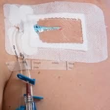
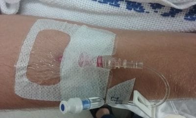
4. Apply a sterile dressing. Dressings can be transparent plastic type, or sterile gauze used with good quality, preferably sterile, tape; but they must be sterile (Marsh et al. 2015, Ullman et al. 2015). Choose a dressing that fits the anatomical location. If the patient is sweating or oozing, plastic dressings will not stick well, and sterile gauze with sterile tape is a better choice. While non-sterile paper tape seems to be everywhere in hospitals, it's not ideal as it adheres poorly, is rough on skin, and is non-sterile. Better quality tapes provide stronger, gentler adherance to skin and stay in place longer. Sterile tape should be sought, espcially if placed close to the wound (ask your IV-starter kit provider to include sterilised tape). Try silicone dressings and tapes for patients whose skin may tear or is irritated. Don't compromise your sterile dressing by placing non-sterile foam under the hub to reduce pressure, or use non sterile tape under the dressing - other sterile foam and tape are available. Patients with poor skin benefit from skin protectant barrier solution applied before the dressing. Of course, let this dry before applying the dressing.
5. No tension! Don't stretch plastic dressings as you apply them, or place dressings/tape so that skin is pulled away from its natural resting shape. It can be tricky to stop some dressings sticking to themselves once you've removed the backing, but don't hold them in a stretched manner as you place them on the skin. Dressings need to be placed gently in a direct downwards motion onto the skin. There should be no tension on the skin in any direction, as this causes skin injury and encourages the dressing to become loose. Gently pat rather than smooth the dressing into place, so there are no gaps or wrinkles, and to activate the adhesive across the entire dressing. Further, don't wrap tape tightly all around the limb. IV devices can fail due to the torniquet-like effect of too much tape. Sure, it's good to keep the line in, but it's not much use if it's blocked off, and it's pretty uncomfortable for the patient too.
6. Apply both a dressing AND a securement. Way back when, we all thought dressings were enough to keep the wound clean, as welll as to hold the IV in place. It's clear now that most dressings need additional securement to hold the IV and any attached tubing to the skin. This extra securement of the catheter prevents not just the line falling out of the vein, but also 'micro-movement' of the device within the vein. The cells inside the vein are delicate - even the smallest IV movement causes irritation, pain, thrombosis, and swelling of the vein. The less micro-motion the less chance the IV will be occluded or 'tissued' when you want to use it.
There are many ways to get additional securement for different IV types. These include two-part dressings which hold the device both from above and below, additional 'securement devices' which may stick to the skin or penetrate it with staple-like components, various tapes, sutures, and stretchy bandage tubes (which patients often like). These all reduce the chance of IVs getting caught on tables, bed rails, handles, and so on. One to two drops of medical grade superglue (cyanoacrylate) applied directly to the wound and under the hub on insertion are an effective way to reduce micro-movement, achieve haemostasis in oozy patients, and provide further infection prevention (Bugden et al. 2016, Rickard et al. 2016).
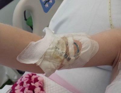
7. If a dressing needs replacing, replace it. Admit it, we've all added layer upon layer of non-sterile paper tape to avoid doing a dressing replacement - sometimes the patient resembles an Egyptian mummy.
But if the dressing has loose edges, has blood under it, or is otherwise dirty, it needs replacing so infections are kept out and the IV kept in. That means cleaning off blood using sterile sodium chloride, re-applying skin decontamination (waiting until it dries), and replacing with a new sterile dressing, and new securement.
8. Remove dressings/tape carefully. When removing, don't rapidly pull these at a vertical angle. This pulls the epidermis, risking skin injury. Instead, loosen the edge of the dressing/tape, and remove 'low and slow' in the direction of hair growth, keeping it close to the skin surface while pulling it back over itself, and supporting the newly exposed skin with your other (gloved) hand. For patients at high risk of skin injury, consider a medical adhesive removal product, but remember these are not sterile, and must be washed off. Each dressing change also requires cleaning the IV wound with sterile saline and reapplying the antiseptic solution. Residue from previous dressings can be removed with medical adhesive remover.
9. Quarterly audit your IV dressings and securements and discuss results with your team. If you walked around your unit today, how many IVs would have dressings that are dry, clean and intact? 100%? How many patients would have skin irritation or injury from dressing/tape use (or misuse)? How many would have pressure tugging on the device from unsecured tubing? How many patients would feel confident that their IV is well secured? This topic can be a simple, regular quality improvement project, leading to local improvements for patients and pride in nursing care standards. Why not run a competition with other wards in your institution as to who has the 'best IV dressings'? Such data could justify more expensive dressing and securement options that actually save money and nursing time through reduced complications. You could present your findings at a nursing conference or write about them in a nursing journal! (Russell et al. 2013, New et al. 2014, Ullman et al. 2016)
10. If you're having trouble, ask a wound and/or vascular access nurse specialist. If a patient has pain, itching, or skin injury from a dressing, or you just can't get the dressings to stay on, seek help from the experts (Ullman et al. 2015). Wound specialists in particular have a deep knowledge of, and access to, the range of specialty dressing products that may be needed in some patients. Even though the wound is from an IV device, it is still a wound and their advice will be invaluable for you and the patient.
References
Bugden, S., K. Shean, M. Scott, G. Mihala, S. Clark, C. Johnstone, J. F. Fraser and C. M. Rickard. 2016. Skin glue reduces the failure rate of emergency department-inserted peripheral intravenous catheters: a randomized controlled trial. Ann Emerg Med 68(2):196-201.
Gorski, L., L. Hadaway, M. E. Hagle, M. McGoldrick, M. Orr and D. Doellman. 2016. Infusion Therapy Standards of Practice. J Infus Nurs 39(Supp1): S1-S159.
Marsh, N., J. Webster, G. Mihala and C. M. Rickard. 2015. Devices and dressings to secure peripheral venous catheters to prevent complications (Review). Cochrane Database Syst Rev(6): CD011070.
Moureau, N. 2013. Safe patient care when using vascular access devices. Br J Nurs 22(2): S14,S16,S18passim.
New, K. A., J. Webster, N. M. Marsh and B. Hewer. 2014. Intravascular device utilisation, management, documentation and complications: a point prevalence survey. Aust Health Rev 38(3): 345-349.
Rickard, C. M., M. Edwards, A. J. Spooner, G. Mihala, N. Marsh, J. Best, T. Wendt, I. Rapchuk, S. Gabriel, B. Thomson, A. Corley and J. F. Fraser. 2016. A 4-arm randomized controlled pilot trial of innovative solutions for jugular central venous access device securement in 221 cardiac surgical patients. J Crit Care 36: 35-42.
Russell, E., R. J. Chan, N. Marsh and K. New. 2014. A point prevalence study of cancer nursing practices for managing intravascular devices in an Australian tertiary cancer center. Eur J Oncol Nurs 18(3):231-5.
Ullman, A. J., M. Cooke, T. Kleidon and C. M. Rickard. 2017. Road map for improvement: point prevalence audit and survey of central venous access devices in paediatric acute care. J Paediatr Child Health 53(2):123-30.
Ullman, A. J., T. Kleidon and C. M. Rickard. 2015. The role of the Vascular Access Nurse Practitioner in developing evidence, promoting evidence-based vascular access practice and improving health services. Vascular Access 1(1): 10-20.
Ullman, A. J., M. Mitchell, F. Lin, K. New, D. Long, M. Cooke and C. M. Rickard. 2015. Dressings and securement devices for central venous catheters (Review). Cochrane Database Syst Rev(9): CD010367.
| Tags:AVATARdressingsintravenous catheterIV managementsecurementvascular access devices |